
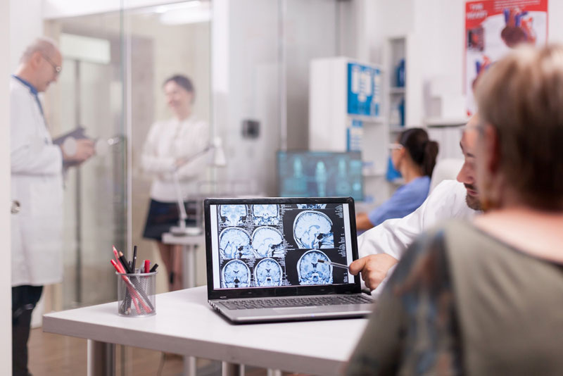
Brain tumours are one of the most complicated neurological diseases and treatment are crucial to maintaining brain function and enhancing the patient’s outcome. Treatment can include surgery, radiation therapy, chemotherapy, targeted therapy, or a combination of these.
The first treatment option is usually surgical removal of the brain tumour, known as excision surgery — it is hoped that the tumour is removed without damaging the normal surrounding brain tissue.
Brain tumour surgery has become safer, more precise, and has a better recovery for patients because of improvements in neuro-navigation, operating microscopes, intraoperative imaging, awake brain surgery and minimally invasive techniques.
India has become a important centre for brain tumour treatment, with neurosurgeons who have been trained abroad, the latest technology, and multidisciplinary teams of neurosurgeons and oncologists, as well as other services to support international patients.
This guide details all the information regarding brain tumour excision surgery, who can benefit from it, available treatment options, and why many international patients come to India for neurosurgery treatment.
A brain tumour diagnosis can be a very stressful experience for patients and families. In addition to the issue of the disease itself, people often have concerns about the effects of treatment on memory, speech, movement, vision or daily activities. Luckily, modern neurosurgery has come a long way and surgeons can now remove many brain tumours with remarkable precision, keeping important neurological functions intact as far as possible.
Surgical removal of a brain tumour may be the treatment’s first and most crucial phase for both benign and malignant brain tumours. The main goals are to remove as much of the tumour as safely as possible, to relieve the pressure on the brain, to help the neurological symptoms, provide tissue for an accurate diagnosis and support further treatment (if needed).
International patients looking for the best neurosurgery care can find world class hospitals in India with the latest technology, highly experienced neurosurgeons, extensive rehabilitation programs and support services for international patients, all at a fraction of the cost of what’s available in many developed countries.
Gliomas are tumours that arise from cells known as glia, which help to sustain and protect nerve cells. They can develop slowly or rapidly, and usually require a surgical procedure for removal followed by further treatment depending upon the grade.
Meningiomas grow from the protective membranes that cover the brain (meninges). While most are benign and slow-growing, larger tumours may compress brain tissue nearby, and need to be surgically removed.
These tumours occur in the pituitary gland and can impact its function in making hormones or sight or both. Numerous can be retrieved using minimally invasive endoscopic surgery via the nose.
Tumour of the nerve associated with hearing and balance. Patients can suffer from hearing loss, tinnitus, or dizziness or loss of balance.
These tumours can be found in the brain as a secondary location to a cancer that has developed in another part of the body, such as the lung, breast, kidney or skin (melanoma). Treatment consists of surgery and either radiation or systemic cancer treatment.
A tumour derived from the astrocytes, a type of glial cell. These tumours can be low-grade and slow-growing, as well as very aggressive.
A relatively rare brain tumour which originates from the oligodendrocytes and tends to grow slowly. Typical treatment involves surgery and may be combined with radiation therapy or chemotherapy.
These tumours develop from the cells that line the ventricles of the brain or the spinal canal and can cause blockage of the fluid which circulates in the ventricles, so they need to be removed by surgery.
A benign tumour in an unusual place near the pituitary gland. Its proximity to brain areas responsible for vision, hormones and growth makes it a potential to affect the vision, hormones and growth.
A rapidly growing tumour that mainly occurs in children but can also develop in adults. Typically treated with surgery, radiation and chemotherapy.
For many, it is not fully understood what causes a brain tumour. But there are some factors that can make it easier to get some kinds of brain tumours.
Brain tumours have a higher risk in some inherited disorders like Neurofibromatosis, Li-Fraumeni syndrome and Turcot syndrome.
People who’ve had radiation treatment to the head for other health problems might be a little higher risk for brain tumours later in life.
While the majority of brain tumours are not inherited, a few can develop in families that have certain genetic syndromes.
There are certain tumours of the brain that occur more often in older adults and others that are often seen in childhood.
Other cancers, like lung cancer, breast cancer, kidney cancer or skin cancer can spread to the brain and create secondary brain tumours.
The cause in most patients is never identified and brain tumours can develop in the absence of any identifiable risks.
Brain Tumour Excision Surgery is an advanced neurosurgical procedure which is used to remove all or a portion of the brain tumour while sparing as much brain tissue and neurological function as possible.
The main aim is not just to remove the tumour, but to ease pressure on the brain, alleviate symptoms, secure tissue for a proper diagnosis and to improve any conditions for further therapy such as radiation, chemotherapy or targeted therapy, if necessary. It is not possible to remove all brain tumours.
Depending on location, size, relationship to critical brain structure and the type of tumour (benign or malignant), the amount of tumour removal will vary. Today, modern neurosurgical technologies enable doctors to operate safely on patients with tumours that were once difficult or impossible to remove.
The goals of brain tumour surgery are,
The most frequently performed brain tumour surgery is called craniotomy. A small hole is temporarily made in the top of the skull and the neurosurgeon uses microsurgical techniques to reach and remove the tumour. The bone flap is then snugly replaced after the procedure.
Awake brain surgery might be considered for tumours in the brain at the level of speech, movement, memory or vision. In certain phases of the procedure, the patient is awake, able to follow simple commands and the surgical team monitors brain function throughout the surgery. This ensures maximum removal of the tumour without affecting important neurological functions.
Specific tumours, such as pituitary tumours and some skull base tumours, can be treated with an endoscope that is inserted through the nose. This is a less invasive method, which can limit the trauma to the tissue and shorten recovery time, and avoids a large incision.
Tumours at the base of the skull are situated near important nerves and blood vessels and thus surgery must be performed with specially trained techniques. They are often carried out with high-tech microsurgical and endoscopic techniques.
Minimally invasive procedures with smaller incisions and specialized instruments may be indicated for selected patients to minimize blood loss, discomfort and recovery time after surgery.
If a tumour can’t be safely removed at once or if a diagnosis is still not certain, a stereotactic biopsy can be used to take a small sample of tissue but with very precise guidance from the computer and with little impact on other tissue in the brain.
Modern brain tumour surgery is performed using a combination of surgical skill and high levels of technology to ensure accuracy, complete removal of the tumour and prevent damage to important neurological functions.
Neuronavigation systems are similar to GPS for the brain, guiding in real time, using a patient’s MRI and CT imaging. This enables the surgeon to pinpoint the tumour and reduce damage to healthy tissue.
During surgery, intra operative MRI can provide real-time imaging as a way for the surgeon to ensure that the tumour is completely removed before the end of the surgery, which decreases the chances of tumour tissue remaining.
During complex neurosurgical procedures, real-time CT imaging helps to verify the accuracy of the surgery, implant placement (if needed) and anatomical orientation.
High precision surgical microscopes offer high magnification and high light and allow for the tumour removal without damaging fine nerves or blood vessels.
Minimally invasive surgery can be used to access certain types of brain tumours via small openings in the body or natural passages by using endoscopic techniques, which disrupts less tissue and leaves scars that heal more quickly.
Functional mapping is a technique used to identify regions that are involved in speech, limb movement, vision and sensation prior to and during surgical intervention. This is particularly important for tumours in close proximity to important areas of the brain.
Monitoring is used continuously during surgery to detect early changes along nerve pathways, motor function and sensory function and to take appropriate corrective measures to prevent compromise of neurological function.
In some malignant brain tumours, a special dye is used that makes the tumour cells ‘fluoresce’ (glow) under a special microscope placed during surgery. This aids the surgeon in identifying tumour tissue from normal brain and facilitates complete and maximum removal of tumour.
The Cavitron Ultrasonic Surgical Aspirator (CUSA) is an ultrasonic surgical aspiration device that selectively breaks up and removes tumour cells while causing minimal damage to normal brain tissue.
Following surgery, patients are cared for in a Neuro Intensive Care Unit that has the necessary equipment to monitor, manage and support life function continuously and in advance in case of potential complications.
Recovering after surgery is ongoing with individualised rehabilitation plans being incorporated that may involve physiotherapy, occupational therapy, speech therapy, cognitive rehabilitation, nutritional support, psychological counselling and much more to improve functional recovery and quality of life.
Brain tumour surgery recovery is a gradual process, and each patient’s journey depends on the type of tumour, its location, the extent of the surgery performed and whether they need any further treatments like radiation therapy or chemotherapy.
Although many patients benefit with striking improvement of symptoms following surgery, a carefully designed rehabilitation program is essential to the best neurologic and functional recovery.
The procedure is followed by transfer to the Neuro Intensive care Unit (Neuro ICU) where patient is monitored closely by a multi-disciplinary team. The constant supervision helps to identify and treat early complications, and encourages a steady recovery.
At this time, doctors watch for:
The Neuro ICU stay duration varies based on the complexity of surgery and patient condition, and is usually between 24-48 hours.
After stabilisation, patients are treated for further recovery on a regular neurosurgical ward. Medical team members focus on:
The average stay at the hospital would be 5-10 days according to the type of surgery and rate of recovery.
Rehabilitation starts as soon as the patient is able to tolerate it. Early rehabilitations minimise complications, enhance functional recoveries and promote independence.
Depending on indivisual needs and rehabilitation include:
Follow-up includes Clinical neurological exam,
The surgery for the removal of brain tumours is a highly specialized surgery and is considered to be relatively safe, however in the hands of an experienced neurosurgeon. Just as with any big surgery, there are some risks and complications that can arise, however.
Bleeding during and/or after the surgery is a rare but possible complication. Due to advanced surgical technique and continuous monitoring, this is minimised.
Although rare, infections may occur at the surgical wound or within the brain. The risk is kept to a minimum through strict sterilization measures and preventive antibiotics.
Swelling is temporary after the removal of tumours. This is typically treated with drugs and careful supervision of the nervous system.
A few patients may have a seizure prior to or following surgery. Use of anti-seizure drugs is recommended to minimize this risk.
In some cases, the fluid around the brain (CSF) may drain from the surgical area. Treatment can be used to treat most cases successfully.
Depending on where the tumour is located, temporary or permanent weakness, numbness, speech and vision changes, balance or coordination problems may occur. These risks can be substantially decreased by modern brain mapping and neuronavigation.
Frequently, the injury is to blood vessels in the immediate vicinity which could lead to a stroke during or after surgery, although this is rare.
Patients who have a major surgery may get a deep vein thrombosis (DVT) or pulmonary embolism (PE). This risk can be reduced by early mobilisation and prevention.
Some brain tumours can come back, even after surgery. Periodic MRI scans are important for long-term follow-up and early detection and management.
Selecting the right hospital for brain tumour surgery is a very important decision to make. The neurosurgery program provides cutting-edge technology, expertise from highly trained professionals, and compassionate patient care, ensuring that patients receive the best care available.
Our neurosurgeons are very skilled and experienced, specializing in the treatment of both benign and malignant brain tumours, including complex skull base and deep-seated tumours.
A multidisciplinary team including the neurosurgeon, neuro-oncologist, neuroradiologist, radiation oncologist, neurologist, pathologist, rehabilitation specialist and dedicated nurse teams is used for every patient.
Each treatment plan is designed for the patient’s specific type of tumour, where it’s located, neurological condition, age and health, and so on, so that they can receive the best possible treatments and results.
Our specially designed Neuro Intensive Care Unit (NICU) allows for specialized postoperative monitoring, so that any complications can be early detected and managed.
Recovery is not limited to surgery. We provide physiotherapy, occupational therapy, speech therapy, cognitive rehab, nutritional counselling and psychological support, all integrated into the rehabilitation process for maximizing recovery.
Comprehensive support to international patients includes,
No, Brain tumours can be non-cancerous (benign) or cancerous (malignant). But even if a tumour is not cancerous it can still need surgery if it is pressing on other parts of the brain or causing a lot of symptoms.
In general, surgery is indicated if the tumour is causing symptoms, is still growing, is interfering with brain function or if a tissue diagnosis is necessary for additional treatment.
The purpose is to have the greatest possible removal of the tumour while maintaining safety. Complete removal is possible only if the tumour is small, located away from important brain areas, and not attached to them.
The awake craniotomy is a specialized technique indicated for tumours in the vicinity of speech and/or movement centres. The patient is awake for part of the surgery to allow the surgical team to watch important brain functions during the surgery to remove the tumour safely.
The time it takes to have a brain tumour removed varies greatly, from 3 hours up to 8 hours, depending on the size and location of the tumour and the type of surgical procedure used.
Patients will stay an average of 5-10 days in the hospital, but recovery may last longer depending on the patient’s condition and surgery performed.
Depending on the type of the tumour, the nature of the pathological report and the amount of tumour removal, some patients may need more treatment. Discuss with your multidisciplinary team which treatment is most appropriate.
Bleeding, infection, seizures, brain swelling, neurologic deficits, cerebrospinal fluid leak, stroke, and recurrence of tumour are potential risks. Your surgeon will discuss any risks that are associated with your condition prior to surgery.
After consulting with the neurosurgeon, most overseas patients are able to travel once medical clearance has been obtained. Time of recovery, neurological stability and other treatment needs determines the timing.
Yes, sometimes tumours may return after surgery. Follow-up MRIs and consultations with specialists are essential for the early detection and timely management.
This will vary depending on the site of the tumour. When it is possible, advanced techniques like functional brain mapping, neuro-navigation and awake craniotomy reduce the risk of compromising essential brain functions.
Yes, Neurosurgeons provide online consultation for you to review your medical records and receive an expert second opinion prior to traveling.
Physiotherapy, occupational therapy, speech therapy or cognitive rehab can help many patients achieve optimal recovery and regain independence.
There are internationally trained neurosurgeons, state of the art surgical equipment, a multi-specialty team, a comprehensive rehabilitation centre, international patient support and quality treatment facilities at prices that are much lower than in many Western countries, all combined in India.
Excision surgery for brain tumors is a highly specialized procedure that is important to the treatment of both benign and malignant brain tumors. A combination of new technologies in neuro-navigation, microsurgical technique, functional brain mapping, intraoperative imaging and multidisciplinary neuro-oncology care makes it possible to obtain excellent clinical results, while maintaining important neurological functions.
India has emerged as a trusted place for the best brain tumor surgery with a blend of cutting-edge technology, state-of-the-art medical facilities, and personalized patient care. Whether you have a consultation online with one of our experts, arrange your medical visa, have surgery, need recovery support or long-term telemedicine follow-up, our team of experienced professionals have you covered and will give you safe, compassionate, patient-centred care at every step of your treatment journey.
Category : Brain Tumour
